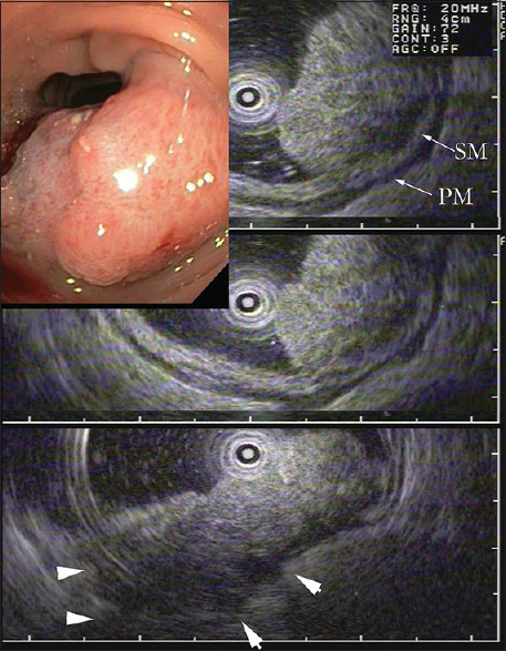
Figure 10.

WLI-colonoscopy: Firm sigmoid polyp, highly suspicious for malignancy. EUS-MP 20 MHz shows a partially intact hyperechoic SM- and PM-layer (upper panel, arrows), but there is also fusion of all layers and hypoechoic invasion beyond the colonic wall corresponding to a tumor category uT3 (lower panel, arrowheads). Histopathology after surgery was pT2 pN0/L0 V0 – G2. A typical case of “overstaging” by EUS-MP probably taking edema for tumor. Nevertheless, the classification ≥ uT2 with indication for surgery was correct
