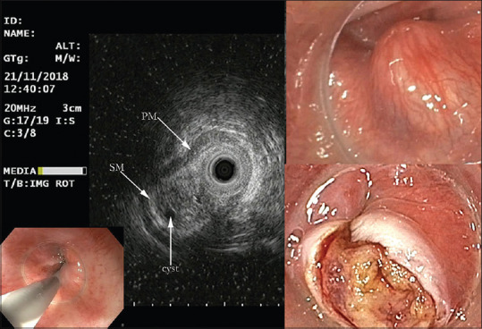
Figure 17.

Submucosal nodule seen from the hypo pharyngeal position in the upper esophageal sphincter. EUS-MP shows submucosal position with intact PM- and SM-layer, heterogeneous echogenicity, small cystic structure. Water filling for optimal imaging was impossible because of the hypo pharyngeal position. Resection was difficult because of firm fibrous attachment; histology was multilocular cyst (benign)
