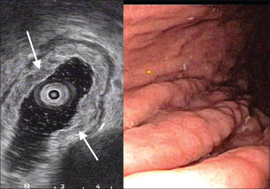
Figure 9.

Apparent mucosal thickening. Infiltration of the LPM and SM of the gastric corpus by slightly inhomogeneous hypoechoic infiltrates (arrows), well separated from PM layer. On white light endoscopy there are slightly edematous gastric folds. Snare biopsy shows mucosa associated lymphoid tissue B-cell lymphoma
