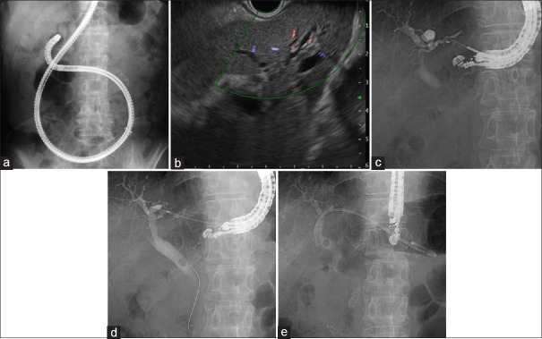
Figure 2.
EUS-guided hepaticogastrostomy for surgically altered anatomy patient with common bile duct stone. (a) Balloon enteroscope was reached into papilla, however biliary selective cannulation was unsuccessful; (b) EUS view of slightly dilated intrahepatic bile duct; (c) the intrahepatic bile duct was punctured using a 22G needle with a 0.018-inch guidewire; (d) the tract was dilated with mechanical dilator. The stone is located on the middle of common bile duct; (e) after exchanging to a 0.025-inch guidewire, the dedicated plastic stent was placed

