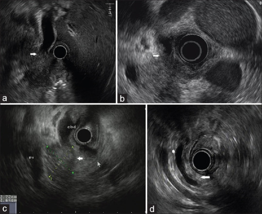
Figure 3.

EUS characteristics of the patient with pancreatic cancer. (a) EUS image showing an irregular hypoechoic mass located in the pancreatic head and a dilated common bile duct which is suddenly interrupted by the mass (arrow). (b) EUS image showing an irregular hypoechoic lesion confined to pancreatic head and an asymmetric thickening of common bile duct wall (arrow). (c) EUS image showing a solitary hypoechoic mass in the pancreatic head with a discernable demarcation between the mass and surrounding parenchyma (wide arrow). (d) EUS image showing an isoechoic pancreatic body/tail (long arrow) and an ectatic main pancreatic duct (short arrow)
