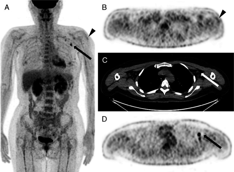
FIGURE 3.

Case 23. FDG PET/CT scan of a 41-year-old woman: 23 days postvaccination. Multiple areas of axillary lymph node accumulation with SUVmax of 5.4 (arrows) and still showing peri-injectional muscle uptake (triangles). A, PET MIP image. B, Axial PET image. C, Axial nonenhanced CT. D, Axial PET image.
