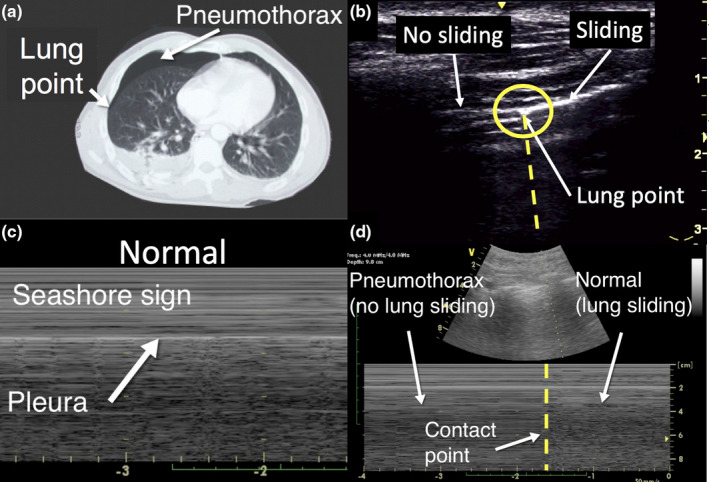
Figure 5.

Pneumothorax. The computed tomography figure (a) of a pneumothorax shows the lung (contact) point of separation of the parietal and visceral pleura. The lung ultrasound image shows the lung point at the intersection of lung sliding on the right and no lung sliding on the left (b). The corresponding lung ultrasound M‐mode image demonstrates lung sliding on the right and no lung sliding on the left of the lung point, a vertical transition zone (d). the lung ultrasound image (c) shows normal lung sliding (seashore sign).
