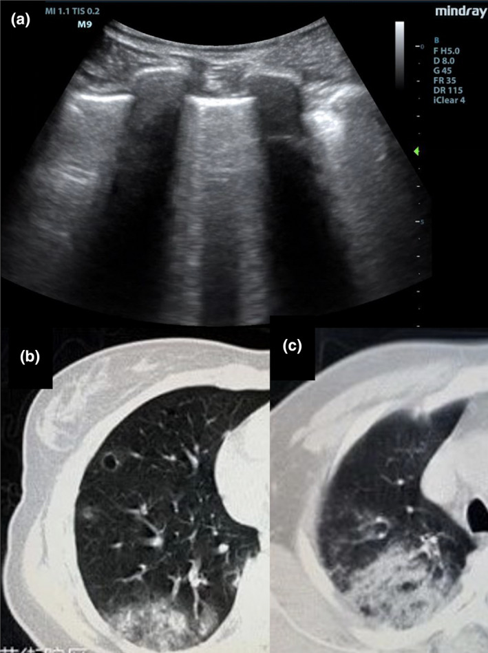
Figure 9.

Lung ultrasound findings in intermediate stage COVID‐19 pneumonia shows thickened and irregular pleura and confluent B‐lines(A). Computed tomography shows ground‐glass opacification in increased quantity, density, and area of distribution (B + C).
