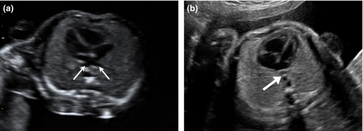
Figure 1.

Four‐chamber View of the Fetal Heart. (a) Normal Pulmonary Venous Drainage. Two Pulmonary Veins are seen Draining into the Left Atrium (Thin Arrows). Note the Distance Between Left Atrium and the Descending Aorta. (b) Abnormal Venous Drainage in Total Anomalous Pulmonary Venous Connection. An Abnormal Vessel is Noted Posterior to the Left Atrium which Represents the Anomalous Pulmonary Venous Confluence (Thick Arrow). The Normal Pulmonary Venous Drainage into the Left Atrium is not Visualised. The Distance Between Left Atrium and Descending Aorta is Increased. The Size of the Left Atrium is Smaller than Usual.
