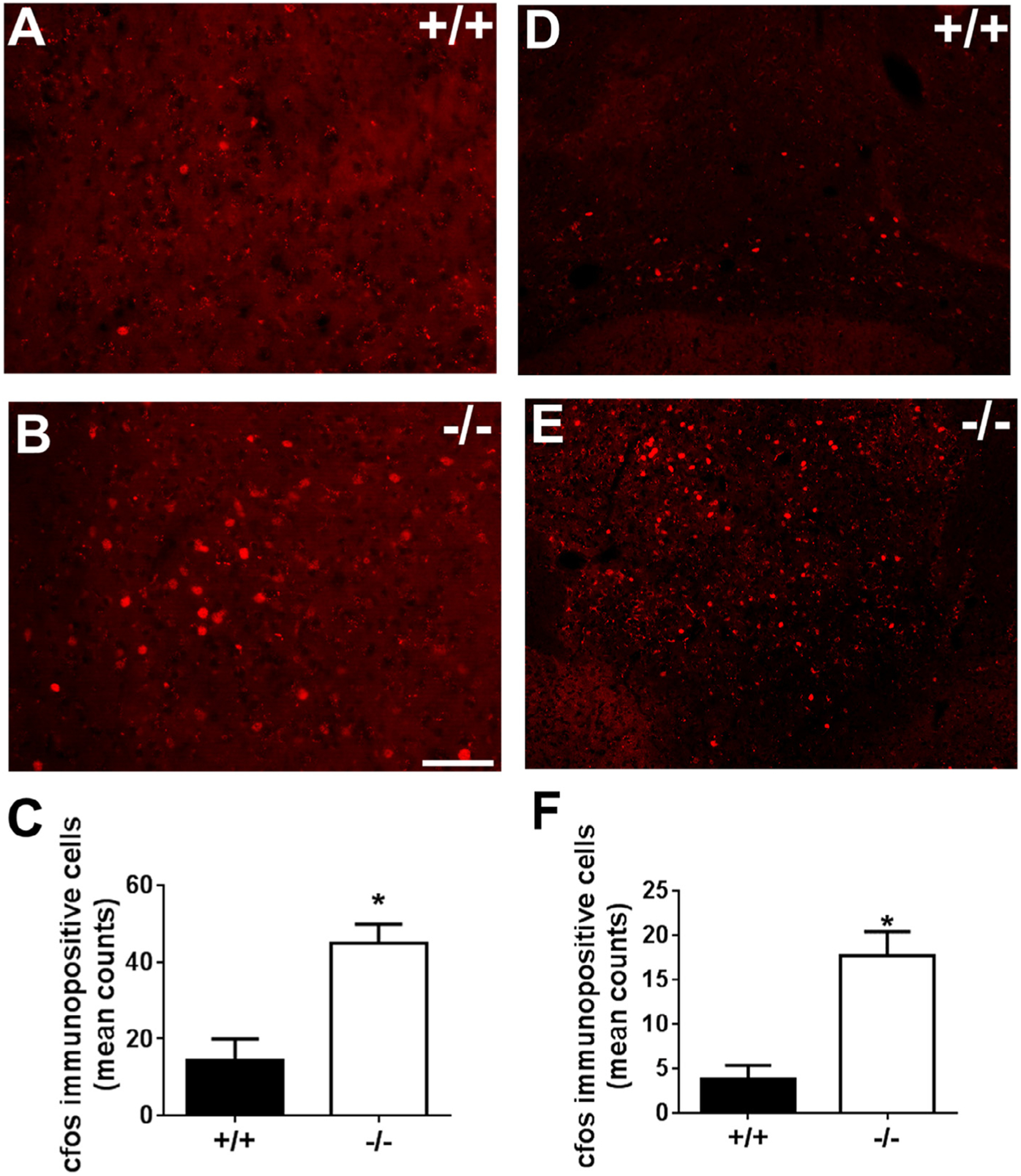
Fig. 6.

cfos-like immunoreactivity in the paraventricular nucleus (PVN) and the ventral tegmental -interpeduncular area (VTA-IPN) of CX3CR1 deficient (−/−) and wild type (+/+) mice. Top panels show representative images of fluorescent cfos immunoreactive nuclei from PVN (A,B) and VTA-IPN (D,E) of +/+ and −/− mice. Bottom panels show bar graphs of quantified data. Significant increase in cfos immunoreactive cells was observed in CX3CR1−/− mice in the PVN (C), and VTA-IPN (F). (*p < 0.05 versus +/+). Data are shown as mean + SEM, n = 6–7/group.
