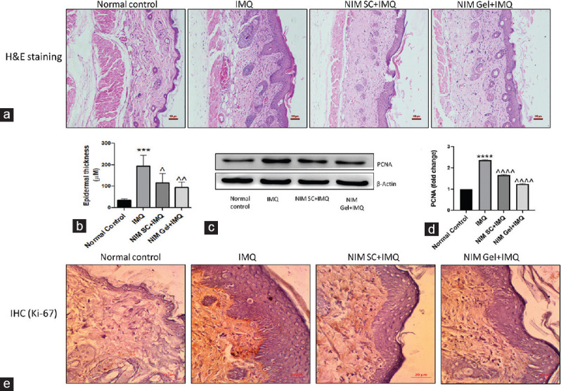
Figure 5.

(a) Representative microphotographs of H and E staining of all experimental groups for evaluating histological changes in skin tissue sections (×10), (b) Graphical representation of epidermal thickness in μM as measured from microphotographs of H and E staining, (c) Western blotting analysis of proliferative protein marker PCNA, (d) Graphical representation of quantified PCNA, (e) Immunohistochemical analysis of another proliferative marker, Ki-67. ****P< 0.0001, ***P< 0.001 of IMQ versus Normal Control; ^^^^P< 0.0001, ^^P< 0.01, ^P< 0.05 of NIM versus IMQ. IMQ = Imiquimod, NIM = Nimbolide, PCNA = Proliferating cell nuclear antigen
