
Figure 4. . Autophagy activity in time course of coronavirus infection.
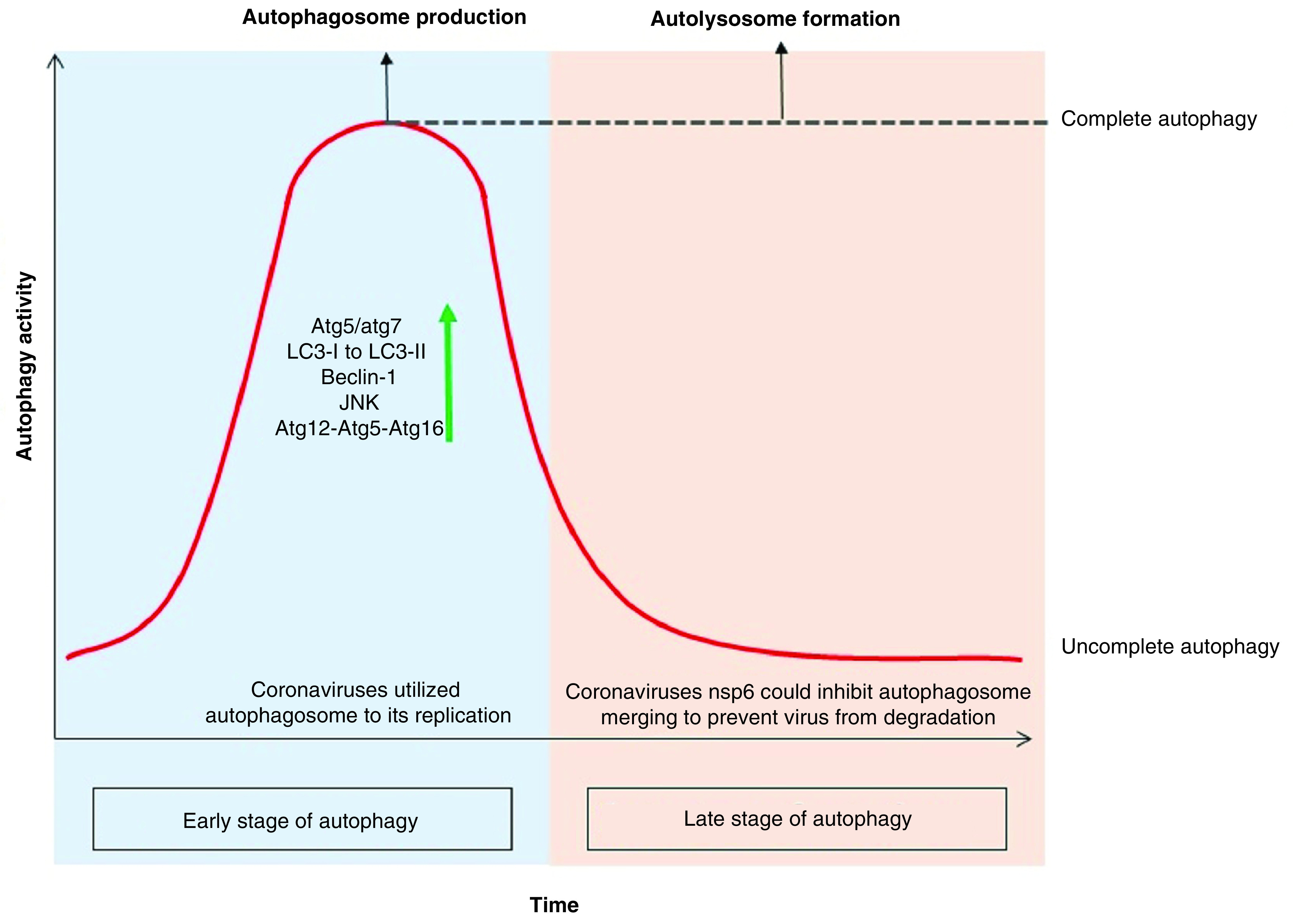
Since the cell infection with the coronavirus, the autophagy activity has been elevating sharply, including an increase in the production of PI3K components like atg12-atg5-atg16 and beclin-1. It also induces the JNK pathway leading to autophagy, which can be induced by promulgate the xeroderma pigmentosum type B splicing of the inositol-requiring enzyme 1 pathway or by a direct effect on the MAPK. According to the studies, deletion of atg5 and atg7 genes reduces coronavirus replication. Coronavirus-infected cells experience increased LC3-I modification due to the addition of glycine to the C-terminal as well as the covalently binding of phosphatidylethanolamine to its amino terminus. This modification results in the production of LC3-II that bind to the outer membrane of the autophagosome and increase the autophagosome marker inner cell. In the late stage of autophagy, coronavirus with the nsp6 infectious protein could constrain autolysosome constructs and turns the cell toward ‘incomplete autophagy’ to keep the virus away from the lysis enzyme of autolysosome.