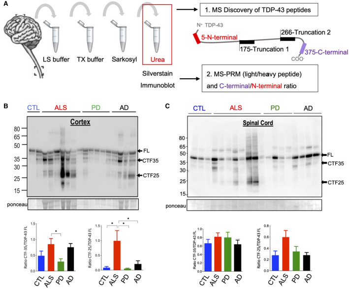
FIGURE 1.

Urea fractionation of brain tissue for the enrichment of pathological TDP‐43. (A) Schematic of sample processing and TDP‐43 peptide identification. Given are the peptides and their initial amino acid within the full‐length TDP‐43 protein. (B and C) Representative immunoblots with an anti‐C‐terminal TDP‐43 antibody recognizing full‐length (FL) TDP‐43 at 43 kDa and smaller C‐terminal fragments (CTFs) of the insoluble (urea) protein fractions from MS‐PRM tissue cohort. (B) Cortex: Quantification of immunoreactive bands at 43 kDa, 35 kDa and 25 kDa demonstrated that the CTF‐35:FL TDP‐43 ratio in ALS was only increased when compared to PD (p = 0.04). CTF‐25:FL TDP‐43 ratio in ALS was increased compared to CTL, PD, and AD (p = 0.03, p = 0.02, and p = 0.048). (C) Spinal cord: Immunoreactive bands showed unaltered CTF‐35:FL and CTF‐25:FL TDP‐43 ratios in ALS compared to CTL, PD, and AD (p = 0.08, p = 0.17, and p = 0.053). Number of cases: 8 CTL, 8 PD, 8 AD, and 15 ALS. One‐way ANOVA with Dunnett’s multiple comparison test
