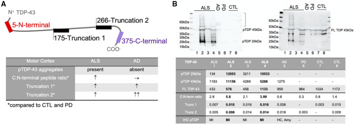
FIGURE 4.

TDP‐43 pathology and peptide quantification in ALS and AD brains. (A) In ALS motor cortices with positive pTDP‐43 pathology an increased C:N‐terminal peptide ratio is present and increased abundance of Truncation 1 and 2 peptides confirms N‐terminal cleavage of TDP‐43. In AD motor cortices with absent pTDP‐43 pathology the C:N‐terminal peptide ratio is normal, but increased Truncation 1 and 2 peptides are indicative of coexisting pathological processing of TDP‐43. (B) Immunoblotting confirms pTDP‐43 immunoreaction (left membrane) at 25 kDa and 45 kDa and a higher molecular smear (]) in ALS motor cortex (MI) with the highest MS‐PRM values for C:N‐terminal ratio, Truncation 1 and 2 and at 45 kDa with a slight higher molecular smear in AD with LATE‐NC in the hippocampus (HC) and Amygdala (Amy) (stage 2). PTDP‐43 is absent in PD and CTL. After stripping FL TDP‐43 at 43 kDa is present in all diagnostic groups (right membrane)
