
FIGURE 2.
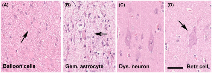
Cytological features of FCD 2 subtypes and its physiologic counterparts. (A) A cluster of balloon cells (arrow) in a 15‐year‐old patient with FCD 2B (H&E staining). (B) A 16‐year‐old patient with focal epilepsy and previous laser ablation. The patient was submitted to a second surgery. Gemistocytic astrocytes were observed in the vicinity of the laser ablation trajectory. (C) Dysmorphic neurons of almost 50 µm diameter seen in a 17‐year‐old patient (same as shown in 2A). (D) This is a giant Betz cell of the primary motor cortex, the Brodmann area 4. These cells resemble dysmorphic neurons. It is imperative, therefore, to know about the surgical resection site and distinguish between occasional norm variants and abundance of abnormally placed and oriented neurons in FCD. Scale bar in D = 50 µm, applies also to A, B, and C