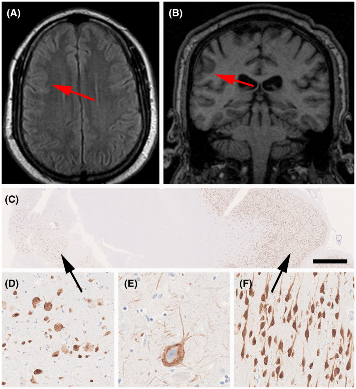
FIGURE 3.

MRI and histopathology findings (see case vignette #1). FLAIR imaging (A) and T1 (B) with red arrows pointing to a subtle lesion with focal cortical thickening and abnormal gyral pattern without hyperintense signal. (C) Low power micrograph of surgical specimen (NeuN immunohistochemistry). The black arrow on the left points to an area of FCD Type 2A, as magnified in (D) and (E) (immunostaining for non‐phosphorylated neurofilament protein). (F) NeuN immunohistochemistry showing well‐oriented pyramidal cells in the adjacent normal‐appearing neocortex (from area highlighted by arrow on the right in 3C). Scale bar in C = 1 mm, in F = 100 µm (applies also to D), in E = 50 µm
