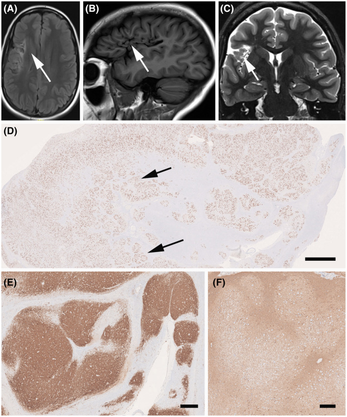
FIGURE 4.

MRI and histopathology findings (see case vignette #2). FLAIR imaging (A), T1 (B), and T2 (C) with encephalomalacia in the right fronto‐parietal operculum and subinsular region, consistent with remote infarction in the right MCA territory (arrows); (D) Low power micrograph of surgical specimen with characteristic neuronal clusters of the cortical architecture (arrows; NeuN immunohistochemistry). MAP2 (E) and GFAP (F) immunohistochemistry revealed the same nodular appearance of the neocortex in FCD Type 3D. Scale bar in D = 1 mm, in E = 100 µm (applies also to D)
