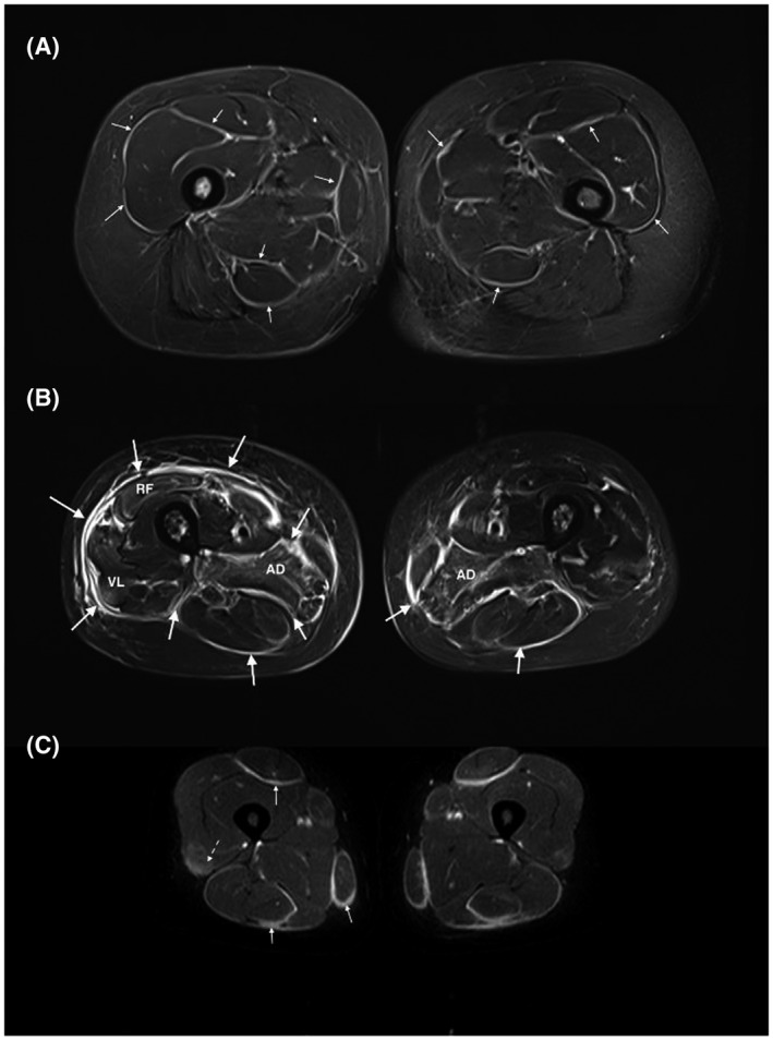
FIGURE 2.

High fascia T2 signals in myositis: (A) patient with an eosinophilic fasciitis displays a diffuse hypersignal of the deep fascia and intermuscular septa (small arrows). The pelvic muscles (i.e., gluteus muscles) are also affected. (B) MRI of a patient with a graft versus host disease involving the fascia (big arrows) (diffuse hyperintensity and thickening of the deep fascia and intermuscular septa) and the muscles (especially both the adductor magnus muscles and the right quadriceps muscle). (C) ASyS patient dysplaying a fasciitis with hyperintense, thickened fascia and intermuscular septa on both sides with symmetrical distribution (full arrows). In addition, presence of a mild myositis attested by a blurred, slight T2 hyperintensity in the quadriceps muscles (dashed arrows). All pictures show thigh muscles MRI images (axial plane, T2 STIR w. seq). RF, rectus femori; VL, vastus lateralis
