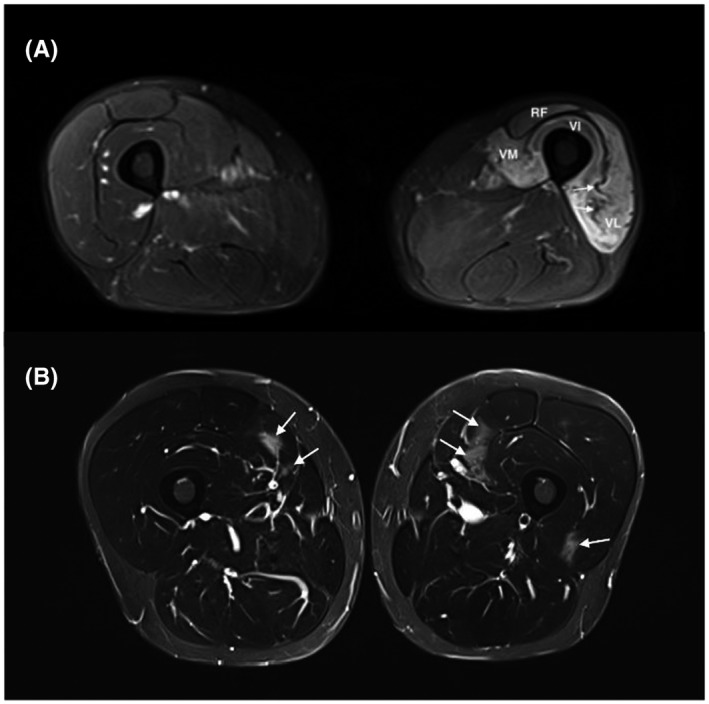
FIGURE 3.

High muscle T2 signals in non‐myositis patients: (A) Neurogenic muscle edema was observed on an MRI performed 4 months after an iatrogenic lesion of the femoral nerve (inguinal hernia surgery). Intense signal and atrophy of the left quadriceps corresponding to the femoral nerve territory are present. Atrophy can be easily identified as compared to the contralateral thigh. Slackness of intramuscular septa (arrow) can also give a clue. (B) MRI 24 h after intense exercise showing patchy areas with increased T2 intensity affecting the quadriceps muscle (arrows), predominantly its medial head, with normal muscle biopsy and spontaneous remission of muscle signs and CK elevation. All pictures show thigh muscles MRI images (axial plane, T2 STIR w. seq). RF, rectus femoris; VI, vastus intermedius; VL, vastus lateralis; VM, vastus medialis
