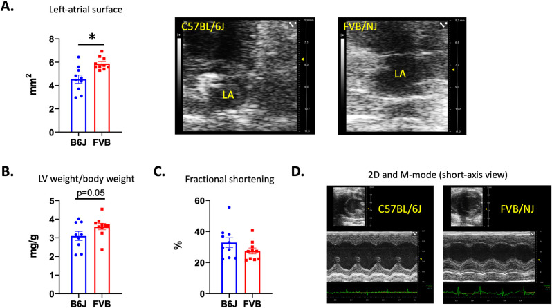
Fig 5. Echocardiographic evaluation showing left cardiac cavities and systolic left ventricular function.
In FVB/NJ mice compared to C57BL/6J echocardiography identified the enlargement of the left atrium (A). Echocardiographic pictures are showing C57BL/6J (upper and left panel) and FVN/NJ (upper and right panel) left atrium obtained in a 2D apical view. An important dilation of the left-atrium is observed in the FVB/NJ mouse. We also observed a left-ventricular hypertrophy (B). At that stage, no reduction of the systolic function was observed (C). In D, the increased thickness of the ventricular walls can be observed on the 2D (upper picture) and the M-mode obtained in a mid-ventricular short axis section of the left ventricle in a C57BL/6J (left) and a FVB/NJ (right). A disorganization of papillary muscles can also be observed in the FVB/NJ mouse case.

