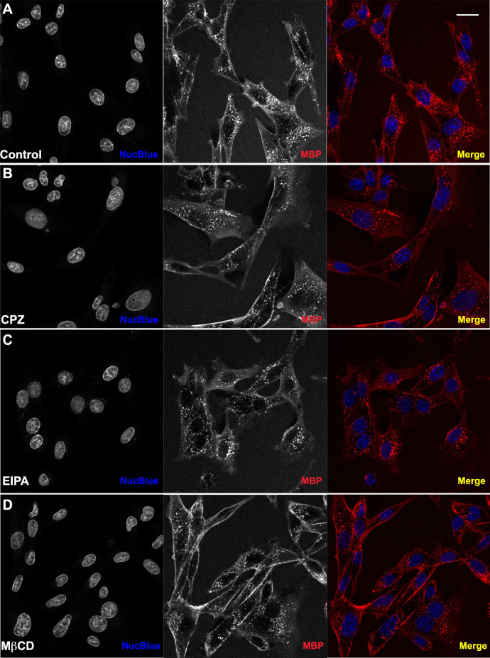
Fig 8. Cell-penetration assays in the presence of inhibitors of endocytosis.
WT TAT-NMR-CaM (1uM) and CBS-MBP (1uM) delivered to BHK21 cells in endpoint static assay (N = 3). Cell nuclei (blue) stained with NucBlue and CBS-MBP (red) labeled with DyLight 650. Cells pretreated with inhibitor for 30 minutes. Proteins introduced to cells in presence of inhibitor. (A) No inhibitor. (B) Chlorpromazine Hydrochloride mediated inhibition of CME. (C) Methyl-β-cyclodextrin mediated inhibition of CvME. (D) 5-(N-Ethyl-N-isopropyl) amiloride mediated inhibition of MP. Cells imaged with 40X objective and scale bar = 20 μm. Red intensity values increased to 45,000 on histogram in Zen Software to improve visualization of uptake.

