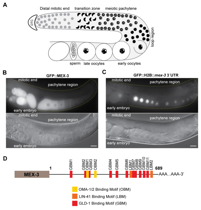
Fig 1. MEX-3 exhibits a unique expression pattern in the germline.
(A) A schematic representing germline organization in C. elegans. One of two gonadal arms is represented. Germ cells undergo mitotic divisions in the distal end and then enter meiosis as they move farther from the distal tip cell. The syncytial meiotic nuclei start to recellularize around the loop region to form oocytes. In the proximal end, late oocytes undergo maturation, get fertilized by the sperm, and then move to the uterus to undergo embryonic development. (B) DIC and fluorescence images of an adult hermaphrodite germline from the strain in which MEX-3 is endogenously tagged with GFP (GFP::MEX-3) [38]. MEX-3 is present in the distal mitotic end, maturing oocytes, and early embryo. (C) DIC and fluorescence images of an adult hermaphrodite germline from the transgenic reporter strain carrying a pan-germline promoter fused to GFP and the mex-3 3´UTR [41]. The reporter is expressed in the distal mitotic end, maturing oocytes, and early embryo. (D) A schematic representing the 3´UTR of mex-3 and its putative binding motifs. Images taken at 40x magnification. Scale bars = 30μm.

