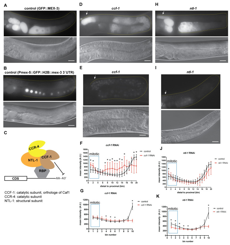
Fig 7. De-adenylation contributes to regulation of MEX-3 in the germline.
(A) DIC and fluorescence images of wild type GFP::MEX-3 animals from the control RNAi. (B) DIC and fluorescence images of the gfp::mex-3 3´UTR transgenic reporter strain from the control RNAi. (C) a model representing the known mechanism of CCF-1, NTL-1, and CCR-4 mediating poly(A) deadenylation. CCF-1 and CCR-4 are the deadenylases while NTL-1 is a structural subunit. CCF-1 is the major de-adenylase. All three proteins are thought to form a complex. (D) DIC and fluorescence images after ccf-1 knockdown in the GFP::MEX-3 strain. GFP::MEX-3 expression was significantly increased in the distal mitotic end. (E) DIC and fluorescence images after ccf-1 knockdown in the gfp::mex-3 3´UTR transgenic reporter strain. (F) quantitative analysis of fluorescence intensity after ccf-1 knockdown in the GFP::MEX-3 strain (n = 15/15). (G) quantitative analysis of fluorescence intensity after ccf-1 knockdown in the transgenic reporter strain (n = 12/12). (H) DIC and fluorescence images after ntl-1 knockdown in the GFP::MEX-3 strain. GFP::MEX-3 expression was significantly increased in the distal mitotic end. (I) DIC and fluorescence images after ntl-1 knockdown in the gfp::mex-3 3´UTR transgenic reporter strain. Transgene expression was not reduced in the distal mitotic end. (J) quantitative analysis of fluorescence intensity after ntl-1 knockdown (n = 13/13) in the GFP::MEX-3. (K) quantitative analysis of fluorescence intensity after ntl-1 knockdown (n = 13/13). (*) indicates statistical significance, adjusted p-value ≤ 0.05. All p-values for this figure are reported in S7 and S8 Tables. Scale bar = 30 μm.

