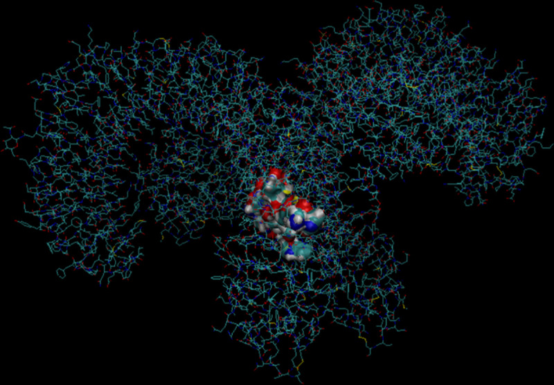
Fig 3. 3D model of Pediocin A1 interacting with TLR-4 produced using the.pdb file imported from RCSB PDB to VMD.

TLR-4 has been drawn in lines in order to distinguish between Pediocin A1 and TLR-4. Like microcin, pediocin appears mainly globular in shape, the TLR-4 is arranged as a heterodimer of two chains converging in the centre, Pediocin A1 interacts with both chains in the middle of this convergence [27].
