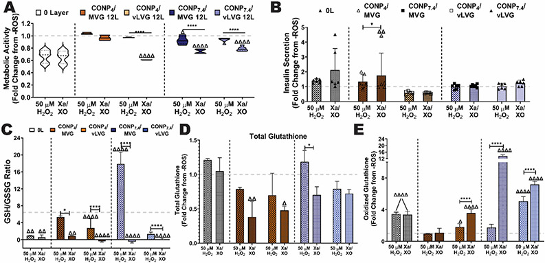
Figure 9. Protection of encapsulated beta cells from exogenous ROS insults was dependent on coating material properties.
The impact of oxidative stress from either H2O2 or SO on the underlying cells was evaluated and summarized as fold change from untreated controls (grey dashed line). Metabolic activity (A) and insulin secretory response to a glucose challenge (B) of encapsulated cells following treatment with H2O2 or Xa/XO (SO generating). Uncoated or microbeads coated with 12-layers of the designated coating formulations were tested. The effect of 12-L coatings on protecting cells was also measured via reduced:oxidized glutathione (GSH:GSSG) ratio (C), total glutathione (D), and oxidized glutathione (E). Grey dashed line: untreated controls. Two-way and one-way ANOVA with Tukey's post-hoc test: ****p < 0.0001, ***p < 0.001, **p < 0.01, and *p < 0.05, where * represents differences between groups, Δ compares 12-L coatings to 0-L beads, and γ compares groups with same alginate MW and varying pH of CONP.

