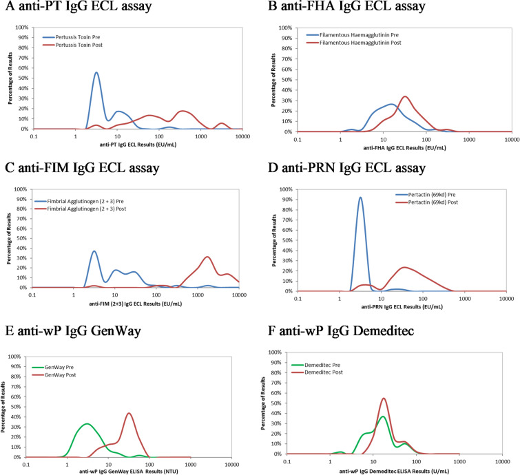
Fig. 6.
Pre- and post-vaccination antibody concentration distribution for ECL assay (anti-FHA, anti-PT, anti-FIM, and anti-PRN), GenWay (anti-wP) assay, and Demeditec (anti-wP) assay. (A) anti-PT IgG ECL (EU/mL), (B) anti-FHA IgG ECL (EU/mL), (C) anti-FIM IgG ECL (EU/mL), (D) anti-PRN IgG ECL (EU/mL), (E) anti-wP IgG GenWay ELISA (NTU) and 6F anti-wP IgG Demeditec ELISA (Units/mL). The pre-vaccination concentration distribution is shown in blue for the ECL methods or green for the anti-wP ELISAs and the post-vaccination concentration distribution is shown in red for all

