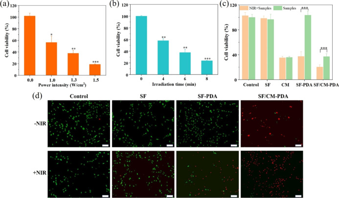
Figure 7.
Cell viability of MG-63 cells after being incubated with the SF-PDA scaffolds under different power densities of NIR irradiation (6 min) (a) and under different irradiation times (1.3 W/cm2) (b). The cell viability of MG-63 cells after being incubated with the SF, SF-PDA, and SF/CM-PDA scaffolds and CM at the same CM concentration level with or without the treatment of the NIR laser (808 nm, 1.3 W/cm2, and 6 min) (c). Live/dead staining images of MG-63 cells incubated with the SF, SF-PDA, and SF/CM-PDA scaffolds with or without the NIR laser (scale bars: 200 μm) (d).

