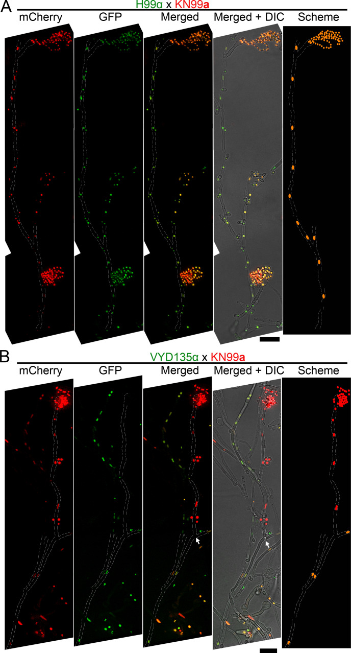
Figure 4. Pan-hyphal microscopy reveals the loss of one parental nucleus during pseudosexual reproduction.
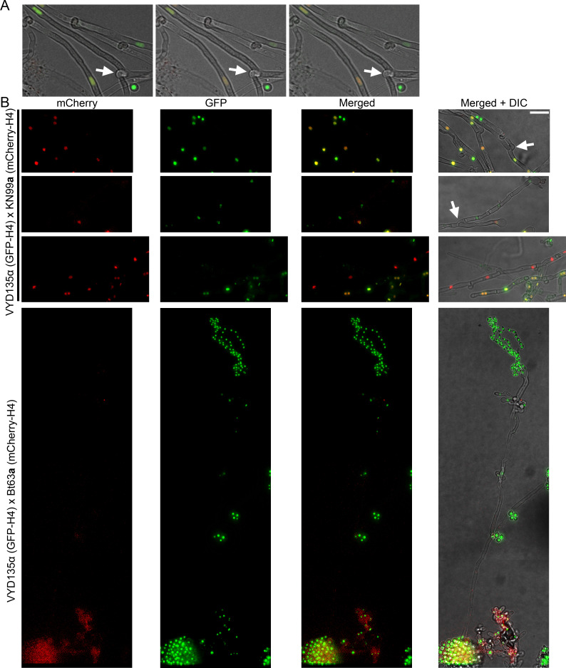
Spore-producing long hyphae were visualized in both (A) wild-type H99α×KN99a and (B) VYD135α×KN99a crosses to study the dynamics of nuclei in hyphae. Both nuclei were present across the hyphal length in the wild-type and resulted in the production of recombinant spores. On the other hand, one of the nuclei was lost during hyphal branching in the VYD135α×KN99a cross and resulted in uniparental nuclear inheritance in the spores that were produced. The arrow in (B) marks the hyphal branching point after which only one of the parental nuclei is present (also see Figure 4—figure supplement 1A). The images were captured as independent sections and assembled to obtain the final presented image. Scale bar, 10 µm.


