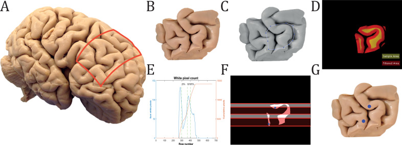
Fig. 1. Sampling of biopsies at BA46.
A Formalin-fixed tissue from three human brains was selected from the brain collection at Aarhus University Hospital. The red box marks the excised area of the tissue. B Fixed coronal block of tissue containing BA46. C Manual delineation of BA46 performed by MATLAB. D Infused image between global threshold image and sample area. The filtered surface of a coronal block of tissue containing BA46 was marked with a red and yellow map that shows the available sample area. E Summation of all white pixels for each row of the binary picture of the sample area. The 1st, 2nd, and 3rd quarter of pixels were marked with a green dashed line. F The two biopsies can only be sampled in either the 1st and 3rd quarter(white area) or the 2nd and 4th quarter (red area). In this case, random points in the 2nd and 4th quarters, marked with blue dots, were chosen by the algorithm. G The two chosen biopsies are marked with blue dots on the original block of tissue from (B).

