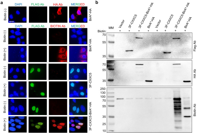
Figure 1.
Biotinylation of endogenous proteins in MCF7 cells. MCF7 cells were transiently transfected for 24 h with an expression vector (pcDNA3.1) bearing the cDNA for 3F-CXXC5, 3F-CXXC5-BirA*-HA, or BirA*-HA. Cells were then treated without (-) or with ( +) biotin (50 μM) and ATP (1 mM) for 16 h. (a) Transiently transfected cells were subjected to immunocytochemistry using the Flag, the HA, or the Biotin antibody followed by an Alexa Fluor 488 (green fluorescein) conjugated goat anti-mouse IgG for the Flag antibody, or an Alexa Fluor 594 (red fluorescein) conjugated goat anti-rabbit IgG for the HA and the Biotin antibody. The scale bar is 20 µm. (b) Total protein extracts of MCF7 cells were subjected to SDS-10%PAGE followed by WB using the Flag, the HA, or the Biotin antibody followed by an HRP-conjugated goat-anti mouse secondary antibody for the Flag (Advansta R-05071–500) or goat-anti-rabbit secondary antibody for the HA or the Biotin antibody (Advansta R-05072–500). Molecular masses (MM) in kDa are indicated.

