Figure 4.
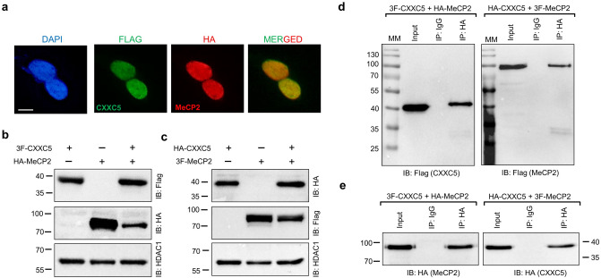
Interaction of CXXC5 and MeCP2. (a) The intracellular localization of CXXC5 and MeCP2 when co-synthesized is assessed with ICC of HEK293 cells transiently transfected for 48 h with the expression vector bearing the 3F-CXXC5 or the HA-MeCP2 cDNA. The Flag (green channel) or the HA (red channel) antibody was used to detect 3F-CXXC5 and HA-MeCP2, respectively. DAPI was used for DNA staining. The scale bar is 10 μm. (b,c) To examine the co-synthesis of CXXC5 and MeCP2, nuclear extracts of HEK293 cells transiently transfected with the expression vector bearing (b) the 3F-CXXC5 and/or the HA-MeCP2 or (c) HA-CXXC5 and/or the 3F-MeCP2 cDNA were subjected to WB analysis. Proteins were immunoblotted (IB) with the Flag or HA antibody. HDAC1 used as a loading control was probed with the HDAC1 antibody. (d,e) The nuclear extracts of HEK293 cells co-synthesizing 3F-CXXC5 and HA-MeCP2, or co-synthesizing HA-CXXC5 and 3F-MeCP2 were subjected to Co-IP using the HA antibody or the isotype-matched IgG followed by immunoblotting using (d) the Flag or (e) the HA antibody. Molecular masses (MM) in kDa are indicated.

