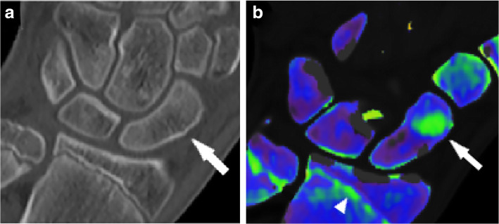
Fig. 4.
Hand dual-energy computed tomography scan in a patient with a non-displaced scaphoid fracture, which was confirmed at magnetic resonance imaging (not shown). Standard grayscale series (a) shows a subtle cortical interruption, which is not clearly suggestive for fracture (arrow). Color-coded virtual non-calcium image (b) depicts the presence of bone marrow edema confirming the hypothesis of a traumatic lesion. Of note, the epiphyseal line on the distal radius and ulna are also color-coded in green (arrowhead). From Dareez et al. [84]

