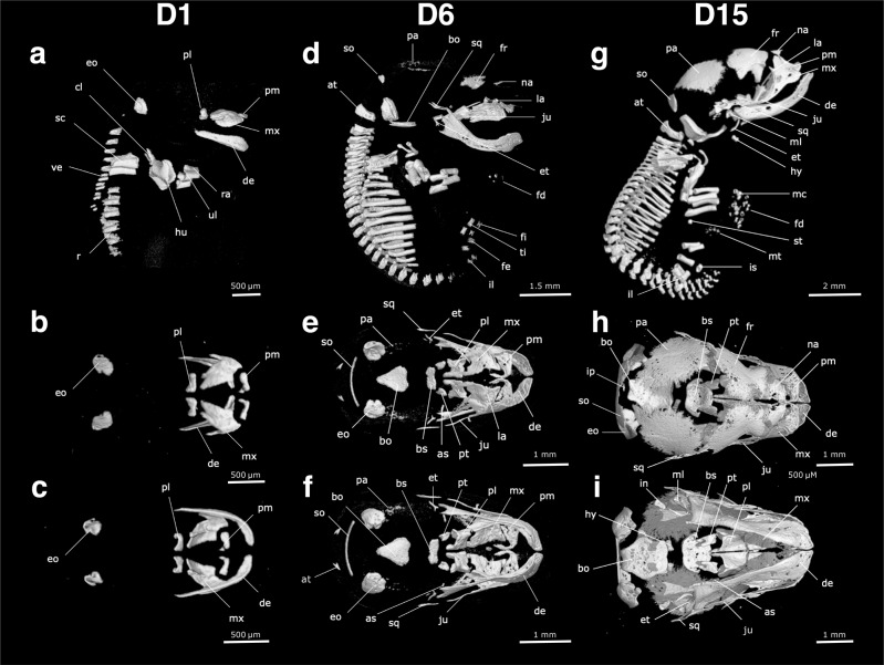
Fig. 4. The skeleton of the fat-tailed dunnart (D1, D6 and D15), as revealed in microCT scans of pouch young.
The right lateral side of the skeleton is shown in (a), (d) and (g), the dorsal view of the skull in (b), (e) and (h), and the ventral view of the skull in (c), (f) and (i). Pouch young one day after birth (D1) are shown in (a), (b) and (c). Pouch young six days (D6) after birth are shown in (d), (e) and (f). Pouch young 15 days after birth (D15) are shown in (g), (h) and (i). as = alisphenoid, at = atlas, bo = basioccipital, bs = basisphenoid, cl = clavicle, de = dentary, eo = exooccipital, et = ectotympanic, fd = forelimb digits, fe = femur, fi = fibula, fr = frontal, hu = humerus, hy = hyoid, in = incus, il = ilium, ip = interparietal, is = ischium, ju = jugal, la = acrimal, mc = metacarpals, ml = malleus, mt = metatarsals, mx = maxilla, na = nasal, pa = parietal, pl = palatine process, pm = premaxilla, pt = pterygoid, ra = radius, ri = ribs, sc = scapula, so = supraoccipital, sq = squamosal, st = sternum, ti = tibia, ul = ulna, ve = vertebrae.

