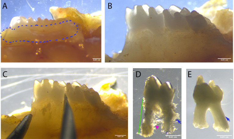
Figure 1. Dissection procedure.

A. Dissect the maxilla and mandible and remove the soft tissue around the tooth (circled area). B. Make sure there is no soft tissue left around the teeth. C. If the tooth is hard to extract, place a needle between the teeth and move around to loosen the tooth, then attempt to extract again. D. The extracted tooth, before enzyme digestion, should show PDL tissue on the root surface (blue arrow); make sure there is no bone tissue in the furcation area (magenta triangle). Keep only the intact teeth (green lines). E. Extracted teeth show a smooth root surface with no PDL tissue attached.
