
Figure 3. Image of HE and immunohistochemical stained section of tumors and testis after transplantation of iPSCs.
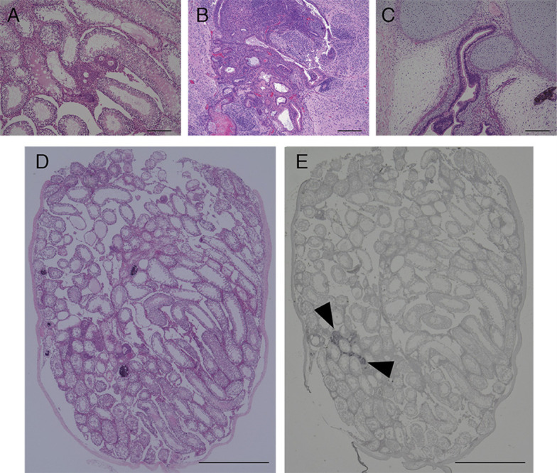
HE staining section of tumors and testis of mice transplanted with NMR iPSCs 10 weeks after transplantation (A and D), mouse iPSCs 4 weeks after transplantation (B) or human iPSCs 10 weeks after transplantation (C). Immunohistochemical analysis of GFP (E). Arrowheads indicate the transplanted GFP-labeled NMR iPSCs. Scale bar, 200 µm (A-C) and 1 cm (D and E).