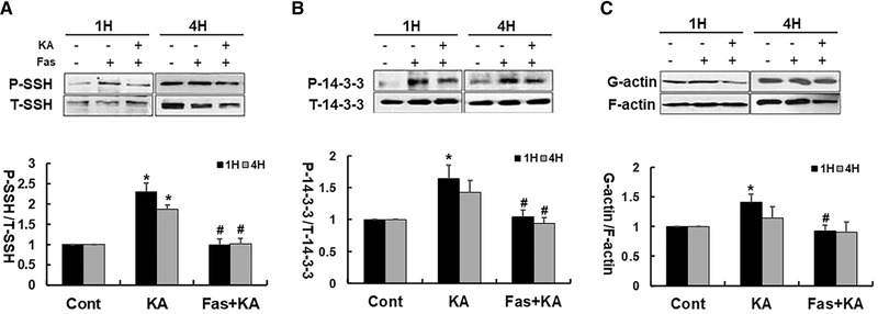
FIGURE 4.

Fasudil significantly downregulated the KA‐induced increase of P‐SSH and P‐14‐3‐3 protein in Neuro‐2a cells. (A) Top: Representative western blots of P‐SSH1 and T‐SSH1 protein. Bottom: Quantification of P‐SSH1 protein expression. (B) Top: Representative western blots of P‐14‐3‐3 and T‐SSH1 protein. Bottom: Quantification of P‐14‐3‐3 protein expression. (C) Top: Representative western blot for actin (G‐actin and F‐actin). Bottom: Summarized data for all experiments show that KA caused markedly decrease of filamentous actin 1 hour after KA treatment, and fasudil alleviated KA‐induced actin depolymerization. *p < .05 by one‐way ANOVA compared to control group (n = 5). #p < .05 by one‐way ANOVA compared to KA‐treated group (n = 5)
