FIGURE 6.

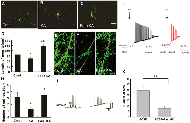
Fasudil pretreatment rescued KA‐induced neurite outgrowth inhibition and spine loss in primary cultured hippocampal neurons. (A–C) The representative rhodamine‐phalloidin image of primary cultured hippocampal neurons at DIV 7 days. Scale bar = 50 μm. (D) Quantification of the neurite length in primary cultured hippocampal neurons *p < .05 by one‐way ANOVA compared to control group (n = 20). #p < .05 by one‐way ANOVA compared to KA‐treated group (n = 20). (E–G) Presentative rhodamine‐phalloidin images of spines on primary cultured hippocampal neurons at DIV 14 days. Scale bar = 10 μm. (H) Quantification of the number of spines in primary cultured hippocampal neurons in different conditions. (I) Presentative trace showed the seizure‐like activity on primary cultured hippocampal neurons evoked by KA application. (J) Presentative trace of the AP with or without fasudil before KA application on hippocampal slices. (H) Quantification of numbers of AP with or without fasudil before KA application on hippocampal slices. *p < .05 by one‐way ANOVA compared to control group (n = 6). # p < .05 by one‐way ANOVA compared to KA‐treated group (n = 6)
