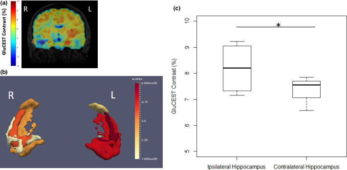
FIGURE 1.

Increased 3‐D GluCEST signal in the ipsilateral hippocampus in a 26‐year‐old with MRI‐negative left temporal lobe epilepsy. (a) Coronal slice of full‐brain GluCEST Map registered to MPRAGE. All coronal slices were averaged across 5 voxels in each slice to improve SNR and to mimic a thick slab (5 mm) from the 2D sequence, with GluCEST contrast percentage scaling from −4 (blue) to 10 (red). (b) After masking the GluCEST map to the hippocampus using T2‐based ASHS hippocampal segmentation, the GluCEST signal in each voxel, averaged across all 4 patients, was averaged into 6‐quantiles (for better visualization) across hippocampal and extrahippocampal regions of interest, from 1 (yellow) to 6 (red). These were then reconstructed using the Paraview imaging software and scaled appropriately. (c) In 4 left‐sided MRI‐negative temporal lobe epilepsy patients, we see a statistically significant increased GluCEST signal in the ipsilateral (left) hippocampus (p =.048, 1‐tailed 2‐sample paired Student's t test, n = 4), indicating increased glutamate
