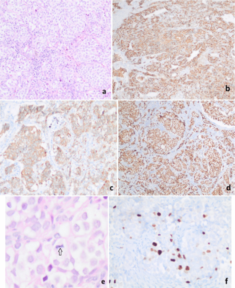
Fig. 4.
Monomorphic epithelial tumor cells forming nests and trabecular structures in the right paratracheal lymph node (H&E, × 200) (a), and immunohistochemically (IHC), diffuse chromogranin staining (IHC, × 100) (b), moderate synaptophysin staining (IHC, × 200) (c), diffuse nuclear TTF-1 staining (IHC, × 100) (d) in the tumor cells. Mitotic figure (black arrow) (H&E, × 1000) (e) and the low Ki-67 labeling index (IHC, × 400) (f) of the neuroendocrine neoplasm are also demonstrated

