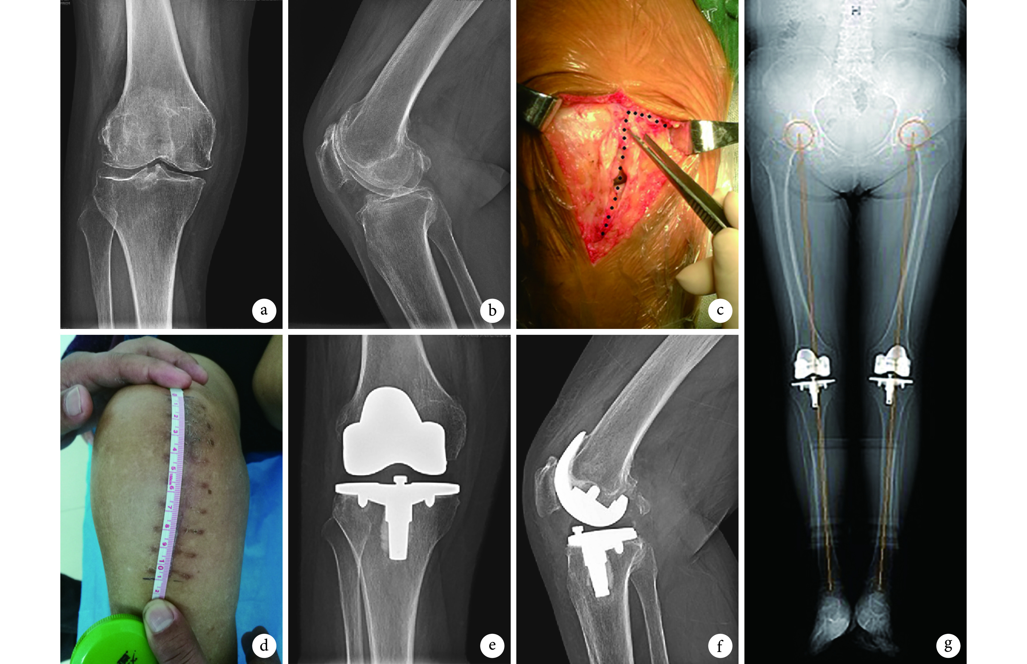
图 1.
A 63-year-old female patient with bilateral knee osteoarthritis underwent TKA via conventional surgical approach for left knee and via mini-subvastus approach for right knee
患者,女,63 岁,双膝骨关节炎行 TKA,左膝行传统入路、右膝行股内侧肌下微创入路
a、b. 术前右膝正侧位 X 线片;c. 右膝术中 L 形(蓝色虚线)切开髌旁内侧腱膜至股内侧肌下缘;d.术后 1 个月右膝切口外观;e、f. 术后 1 个月右膝正侧位 X 线片;g. 术后 5 年双下肢全长 X 线片
a, b. Anteroposterior and lateral X-ray films of the right knee before operation; c. Dissected from medial margin of patella to border of the vastus medialis for deep tissue, like L-shape (blue dotted line) during operation; d. Appearance of the right knee incision at 1 month after operation; e, f. Anteroposterior and lateral X-ray films of right knee at 1 month after operation; g. Full-length X-ray film of bilateral lower extremities at 5 years after operation

