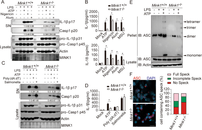
Fig. 1.
Myeloid MINK1 is essential for NLRP3 inflammasome activation. A, B BMDMs from Mink1+/+ and Mink1−/− mice primed with LPS and stimulated with different secondary signals, including ATP, nigericin, aluminum salts (Alum), and monosodium urate crystals (MSU). Supernatants (SN) and cell extracts (lysate) were analyzed using immunoblotting (A). Supernatant IL-1β and IL-18 were analyzed using ELISA (B). C, D BMDMs from Mink1+/+ and Mink1−/− mice primed with LPS and stimulated with ATP, poly (dA:dT), and Salmonella. SN and lysate were analyzed using immunoblotting (C). SN IL-1β was also analyzed using ELISA (D). E, F BMDMs from Mink1+/+ and Mink1−/− mice primed with LPS and stimulated with ATP followed by immunoblotting for ASC oligomerization analysis in cross-linked cytosolic pellets (E). Representative immunofluorescence images and quantification of ASC speck formation are shown in F. *p < 0.05, two-tailed unpaired Student’s t test was used for B and D. The ELISA and western blot results are representative of three independent experiments

