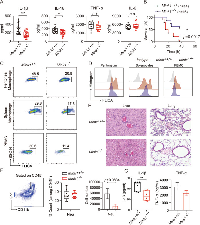
Fig. 2.
MINK1 ablation ameliorates acute inflammation. A Mink1+/+ and Mink1−/− mice were intraperitoneally injected with LPS (25 mg/kg), and IL-1β, IL-18, IL-6, and TNF-α in serum were measured using ELISA after 4 h. Data are the means ± SD (n = 20 mice/group). B The survival rates of Mink1+/+ and Mink1−/− mice injected with LPS (20 mg/kg, n = 14 in Mink1+/+ and n = 16 in Mink1−/−). Surface staining of FLICA was performed on macrophages (gated on F4/80+ cells) from the peritoneal cavity, spleen and blood from Mink1+/+ and Mink1−/− mice to determine the proportion of activated caspase-1 macrophages (C) and the intensity of caspase-1 activation (D). Numbers in or adjacent to outlined areas (or in quadrants) indicate the percentages of cells in each throughout. E Representative histology of liver and lung (hematoxylin and eosin stained) from Mink1−/− and WT mice 4 h after LPS (25 mg/kg) injection. Scale bar represents 100 μm. F, G Mink1+/+ and Mink1−/− mice were intraperitoneally injected with alum (2 mg per mouse), and peritoneal fluid was washed out 6 h later for cell surface staining to determine the percentages and cell number of neutrophils between the two groups. F Data are the means ± SD (n = 6 mice/group). Peritoneal IL-1β and TNF-α levels in Mink1−/− and WT mice were also detected using ELISA (G). *p < 0.05, *p < 0.01, ***p < 0.001, two-tailed unpaired Student’s t test for A, F, and G, and Kaplan–Meier method for mouse survival (B). The ELISA and flow cytometry results are representative of three independent experiments

