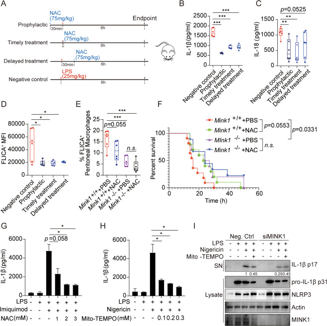
Fig. 6.
NAC significantly alleviates the acute inflammatory response mediated by NLRP3 activation in human and mouse cells. A Schematic diagram of NAC treatment model. B, C Eight-week-old mice were intraperitoneally injected with LPS (25 mg/kg), followed by different NAC treatments at different time points. Serum was collected to detect IL-1β and IL-18. Data are the means ± SD (n= 6 mice/group). D Surface staining of F4/80 and FLICA on macrophages in spleens from C57BL/6 (B6) mice to determine the intensity of caspase-1 activation. E Surface staining of F4/80 and FLICA on peritoneal macrophages from Mink1+/+ and Mink1−/− mice to determine the proportion of activated caspase-1 macrophages. F Survival rate of Mink1+/+ and Mink1−/− mice injected with 20 mg/kg LPS. G, H PBMCs were freshly isolated from healthy donors and LPS-primed PBMCs with NAC or Mito-TEMPO before imiquimod or nigericin stimulation. Supernatant IL-1β was analyzed using ELISA. I LPS-primed THP-1 cells were treated with Mito-TEMPO before nigericin stimulation. Supernatants (SN) and cell extracts (lysate) were analyzed using immunoblotting. *p < 0.05, **p < 0.01, ***p < 0.001. One-way ANOVA was used for B–E, G, and H, and the Kaplan–Meier method was used for mouse survival F. The ELISA and western blot results are representative of three independent experiments

