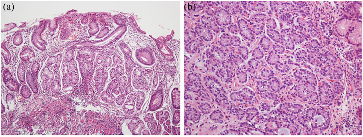
Figure 2.
(a) Histopathological features of autoimmune gastritis (H&E stain, 100×, oxyntic mucosa). As characteristic of autoimmune gastritis, there is absence of parietal and chief cells. These are replaced by intestinal metaplasia and pseudopyloric metaplasia. There is background chronic inflammation. (b) Histopathologic image of type-I gastric neuroendocrine tumor. The tumor cells are well differentiated as evidenced by monomorphic round nuclei. These appear as “nests” infiltrating the lamina propria. Chromogranin A stain could be used to confirm neuroendocrine differentiation (stain not shown). Images are courtesy of Dr M Blanca Piazuelo.

