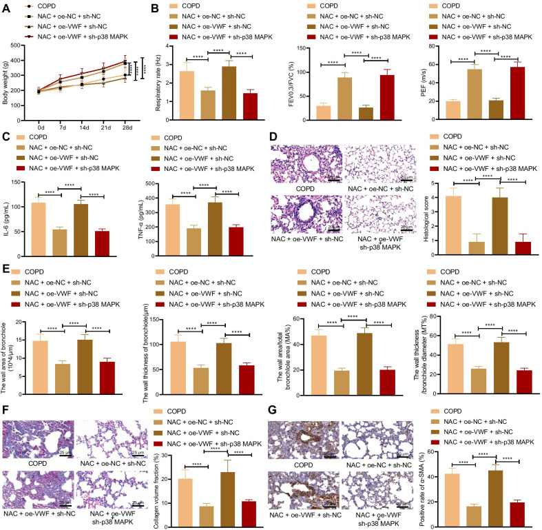
Fig. 7.
NAC relieves pulmonary fibrosis caused by COPD through the VWF/p38 MAPK axis. COPD rats were treated with NAC and oe-VWF and/or sh-p38 MAPK (n = 10). A The weight of COPD rats. B Pulmonary function of COPD rats. C Levels of IL-6 and TNF-α in the serum of COPD rats measured by ELISA. D Histological score of COPD rats detected by HE staining. E Bronchioles area, thickness bronchioles, the wall area/total bronchiole area (MA%) and the wall thickness/bronchiole diameter (MT%) of COPD rats. F Collagen volume fraction in lung tissues of COPD rats detected by Masson’s trichrome stain. G α-SMA level in lung tissues of COPD rats detected by Immunohistochemistry. Cells were treated with CSE and PBS or CSE and NAC. ****p < 0.0001. Data are shown as the mean ± standard deviation of three technical replicates. Data among multiple groups were compared by one-way ANOVA with Tukey’s post hoc test. Data at different time points were compared by two-way ANOVA with Bonferroni post hoc test

