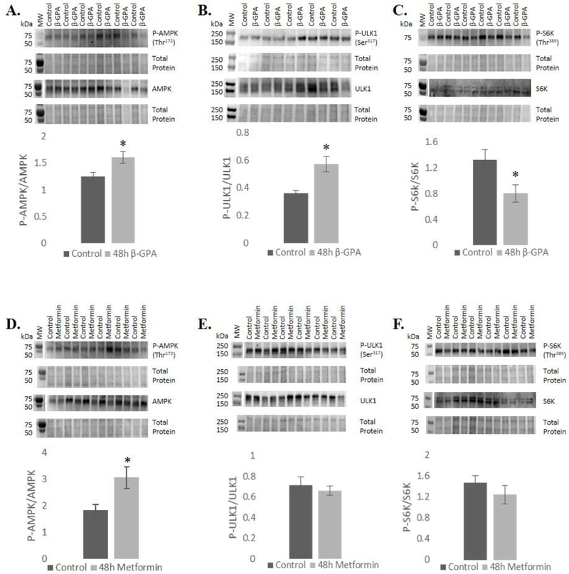
Fig. 2.
48 h β-GPA treatment led to an increase in phosphorylation of A) AMPK, reduced phosphorylation of P70S6K, and increased phosphorylation of C) ULK. Metformin led to increased phosphorylation of D) AMPK, but did not alter phosphorylation of E) P70S6K or F) ULK1. Molecular weight markers typically did not show up in the western blot ECL detection, so in those cases blots were aligned with total protein images and markers from total protein gels are shown adjacent to blots above and separated by a gap. n=6 (n=9 for control and metformin groups of AMPK); *P≤0.05.

