Fig. 3.
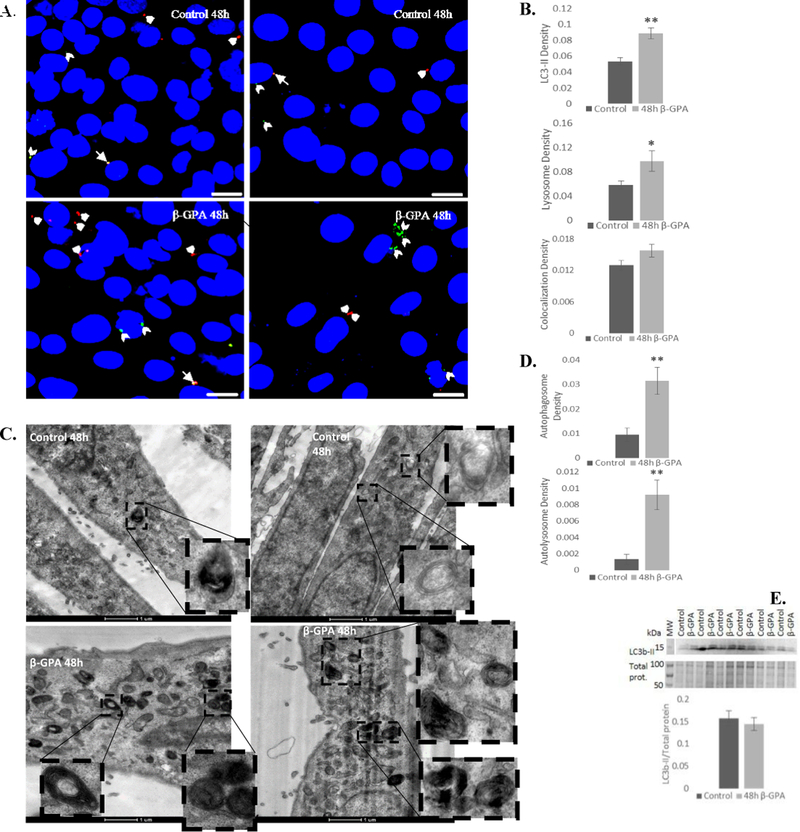
β-GPA enhances autophagy in developing myotubes. A, B) Confocal microscopy revealed that β-GPA treatment led to an increase in autophagosome (green LC3-II labeling; beveled arrowheads) and lysosome (red lysotracker labeling; arrowheads) density, with no significant difference in colocalization (arrows) (n=45). Scale bar = 20 μm. C, D) TEM also revealed that 48h β-GPA increased autophagosome and autolyosome density (n=30). E) 48 h β-GPA treatment did not alter relative expression of LC3b-II. There was insufficient total protein staining near the 15 kDa molecular weight marker, so LC3b-II was normalized to total protein from the 50–100 kDa range. n=6; *P≤0.05, ** P≤0.01.

