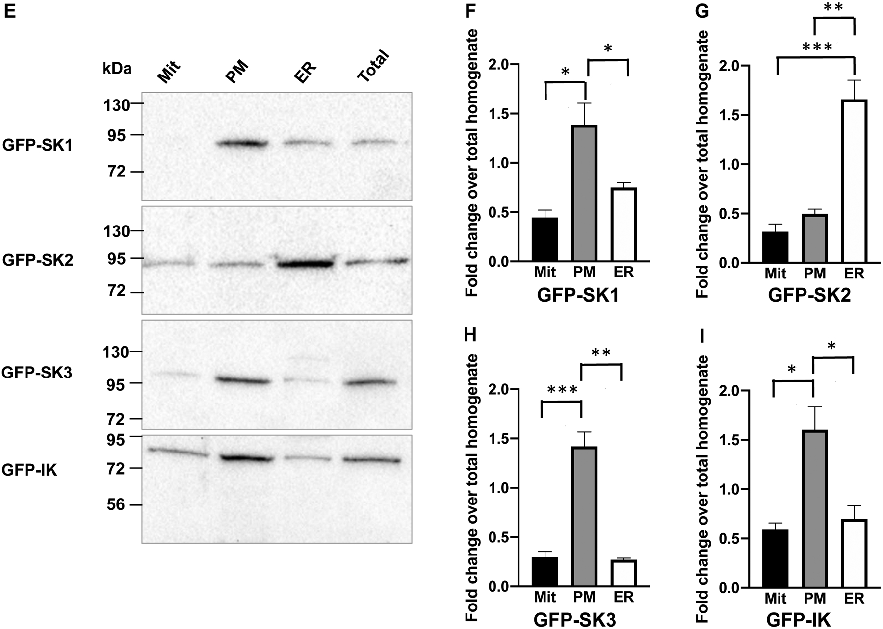
Fig. 3. Subcellular localization of the SK channel subtypes in immortalized endothelial cells.


(A) Cell fractions were verified by immunoblots with antibodies for mitochondria marker (Cytochrome C), ER marker (GRP-78) and plasma membrane marker (Na+-K+ ATPase). In (B), (C) and (D), densitometry results for the subcellular markers are summarized from 3–4 experiments. (E) In cell fractions, the SK2 channel subtype is abundantly expressed on the ER membrane, in contrast to other channel subtypes. In (F), (G), (H) and (I), densitometry results for the SK channel subtypes are summarized from 4–6 experiments. All data are shown as mean ± S.E.M. *P<0.05, **P<0.01, ***P<0.001, Two-way ANOVA followed by Tukey’s post hoc tests.
