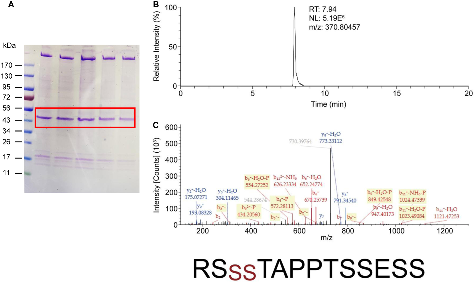
Fig. 5. Nano-LC/MS/MS analyses of SK2 protein isolated from the plasma membrane of the immortalized endothelial cells.

(A). The endothelial cells expressing SK2 channels were subjected to cell fractionation. SK2 channel proteins were purified from the plasma membrane fraction and then separated on SDS-PAGE gel. Protein bands at ~50 kDa and ~17 kDa correspond to SK2 channels (labeled in red box) and calmodulin, respectively. (B). The extracted ion-chromatograms of the peptide “567RSSSTAPPTSSESS580” of SK2 channels (doubly charged ion at m/z 370.80457). (C). The tandem mass spectrum of the peptide was acquired from the doubly charged precursor ion at m/z 370.80457. Fragment ion peaks as b- or y-type ions are also labeled in the spectrum. The peptide sequence including the phosphorylated sites at serines (red color) is indicated at the bottom.
