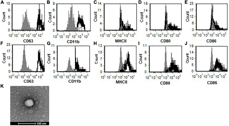
FIGURE 1.
TMEV-infected microglia secrete exosomes that contain activation markers. Exosomes were isolated from uninfected (A–E) or TMEV infected microglia (F–J). The exosomes were labeled with fluorescently labeled antibodies for CD63 (A,F), CD11b (B,G), MHC class II (C,H), CD80 (D,I), and CD86 (E,J). The exosomes were analyzed by flow cytometry for expression of specific markers as shown in the black line compared to isotype control antibodies in the gray line. (K) Isolated exosomes were analyzed by transmission electron microscopy and determined to be 40–80 nm. One representative image is shown. These are representative graphs and images from one experiment of four independent repeated experiments.

